Lab 6 Review Is Onion Skin Multicellular or Unicellular or Colonial
Protista
Protists are any eukaryotic organism that are not an animal, found or fungus. The protists practice non form a natural group, or clade, merely are ofttimes grouped together for convenience. In the popular five-kingdom scheme proposed by Robert Whittaker in 1969, the protists make upwardly a kingdom called Protista, composed of "organisms which are unicellular or unicellular-colonial and which form no tissues. Some protists are meaning parasites of animals (e.grand., five species of the parasitic genus Plasmodium cause malaria in humans and many others cause similar diseases in other vertebrates), plants (the oomycete Phytophthora infestans causes tardily blight in potatoes) or even of other protists. Protist pathogens share many metabolic pathways with their eukaryotic hosts. This makes therapeutic target development extremely difficult - a drug that harms a protist parasite is besides probable to harm its animal/plant host.
The term protista was first used by Ernst Haeckel in 1866. Protists were traditionally subdivided into several groups based on similarities to the "higher" kingdoms such as:
- Protozoa: the unicellular "animal-like" (heterotrophic/parasitic) protozoa which were further sub-divided based on motion such equally (flagellated) Flagellata, (ciliated) Ciliophora (or Ciliata), (phagocytic) amoeba and spore-forming Sporozoans
- Protophyta: the "found-like" (autotrophic) protophyta (mostly unicellular algae)
- Molds: the "fungus-like" (saprophytic) slime molds and water molds.
The taxonomy of protists is always changing. Newer classifications attempt to present monophyletic groups based on morphological (especially ultrastructural), biochemical (chemotaxonomy) and DNA sequence (molecular research) data. However, there are sometimes discordances betwixt molecular and morphological investigations.
View Living Organisms
Amoeba proteus
Amoeba proteus (Figure 14.1) is an amoeba closely related to the giant amoebae. This small protozoan uses tentacular protuberances called pseudopodia to motion and phagocytose smaller unicellular organisms, (which may be greater in size than of amoeba), which are enveloped inside the cell's cytoplasm in a food vacuole, where they are slowly cleaved down by enzymes. It occupies freshwater environments and feeds on other protozoans, algae, rotifers, and even other smaller amoebae. Due to phytochromes, A. proteus may appear in a variety of colors (ofttimes yellowish, dark-green and purple) nether a microscope.
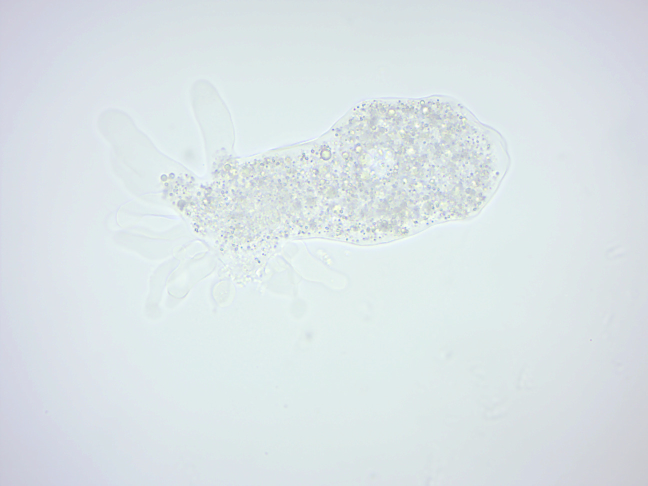
Figure fourteen.1: Amoeba proteus.
Paramecium caudatum
Paramecium caudatum (Effigy 14.ii) is a unicellular, ciliate eukaryote. They can reach 0.25mm in length and are covered with minute hair-like organelles called cilia. The cilia are used in locomotion and feeding. P. caudatum feed on bacteria and small eukaryotic cells, such equally yeast and flagellate algae. In hypotonic conditions (freshwater), the cell absorbs h2o by osmosis. Information technology regulates osmotic pressure with the help of bladder-like contractile vacuoles, gathering internal water through its star-shaped radial canals and expelling the excess through the plasma membrane. When moving through the h2o, they follow a screw path while rotating on the long axis. Paramecium have 2 nuclei (a big macronucleus and a single compact micronucleus). They cannot survive without the macronucleus and cannot reproduce without the micro-nucleus. Like all ciliates, Paramecia reproduce asexually, by binary fission. During reproduction, the macronucleus splits by a blazon of amitosis, and the micronuclei undergo mitosis. The cell then divides transversally, and each new jail cell obtains a re-create of the micronucleus and the macronucleus. Fission may occur every bit office of the normal vegetative cell bicycle. Nether certain conditions, it may be preceded by self-fertilization (autogamy), or it may follow conjugation, a sexual phenomenon in which Paramecia of uniform mating types fuse temporarily and commutation genetic cloth. During conjugation, the micronuclei of each conjugant divide by meiosis and the haploid gametes pass from one cell to the other. The gametes of each organism then fuse to form diploid micronuclei. The onetime macronuclei are destroyed, and new ones are developed from the new micronuclei. Without the rejuvenating effects of autogamy or conjugation a Paramecium ages and dies. Only opposite mating types, or genetically compatible organisms, tin unite in conjugation.

Figure 14.2: Paramecium caudatum.
Euglena
Euglena (Effigy 14.3) is a genus of single-celled flagellate eukaryotes. It is the all-time known and most widely studied member of the grade Euglenoidea, a diverse group containing some 54 genera and at least 800 species. Species of Euglena are plant in fresh and salt waters. They are often arable in quiet inland waters where they may bloom in numbers sufficient to color the surface of ponds and ditches green (E. viridis) or red (E. sanguinea). When feeding equally a heterotroph, Euglena takes in nutrients by osmotrophy, and tin survive without light on a diet of organic matter, such equally beef extract, peptone, acetate, ethanol or carbohydrates. When there is sufficient sunlight for information technology to feed by phototrophy, it uses chloroplasts containing the pigments chlorophyll a and chlorophyll b to produce sugars by photosynthesis. Euglena'southward chloroplasts are surrounded by three membranes, while those of plants and the dark-green algae (amid which earlier taxonomists ofttimes placed Euglena) take simply 2 membranes. This fact has been taken as morphological bear witness that Euglena's chloroplasts evolved from a eukaryotic light-green alga. Thus, the intriguing similarities betwixt Euglena and the plants would have arisen not because of kinship just because of a secondary endosymbiosis. Molecular phylogenetic assay has lent support to this hypothesis, and it is now generally accustomed.
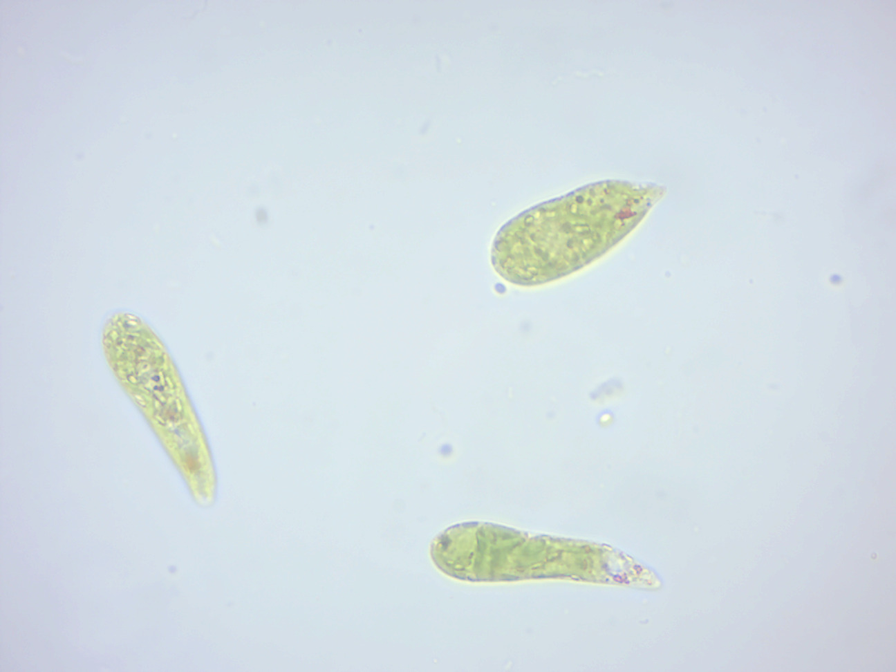
Figure xiv.three: Euglena.
Peranema
Peranema (Figure xiv.4) is a genus of free-living flagellate, with more 20 accepted species, varying in size betwixt 8 and 200 micrometers. They are institute in freshwater lakes, ponds and ditches, and are often abundant at the lesser of stagnant pools rich in decaying organic material. Although they belong to the grade Euglenoidea, and are morphologically similar to the green Euglena, Peranema have no chloroplasts, and cannot feed by autotrophy. Instead, they capture alive prey, such as yeast, leaner and other flagellates, consuming them with the assist of a rigid feeding apparatus called a "rod-organ." Unlike the light-green Euglenids, they lack both an eyespot (stigma), and the paraflagellar trunk (photoreceptor) that is ordinarily coupled with that organelle. Nevertheless, while Peranema lack a localized photoreceptor, they do possess the light-sensitive protein rhodopsin, and reply to changes in light with a characteristic "crimper behavior."
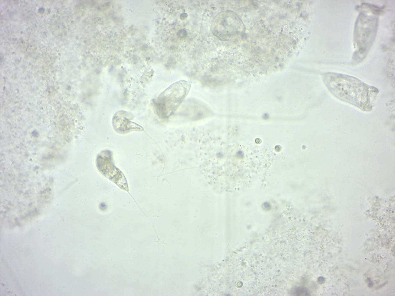
Figure 14.iv: Peranema.
Chlamydomonas
Chlamydomonas (Figure 14.5) is a genus of green algae consisting of unicellular flagellates, found in stagnant water and on damp soil, in freshwater, seawater, and even in snowfall as "snow algae". Chlamydomonas is used equally a model organism for molecular biological science, particularly studies of flagellar motility and chloroplast dynamics, biogeneses, and genetics. 1 of the many striking features of Chlamydomonas is that it contains ion channels, (channelrhodopsins), that are direct activated by light. These proteins are used in optogenetics.
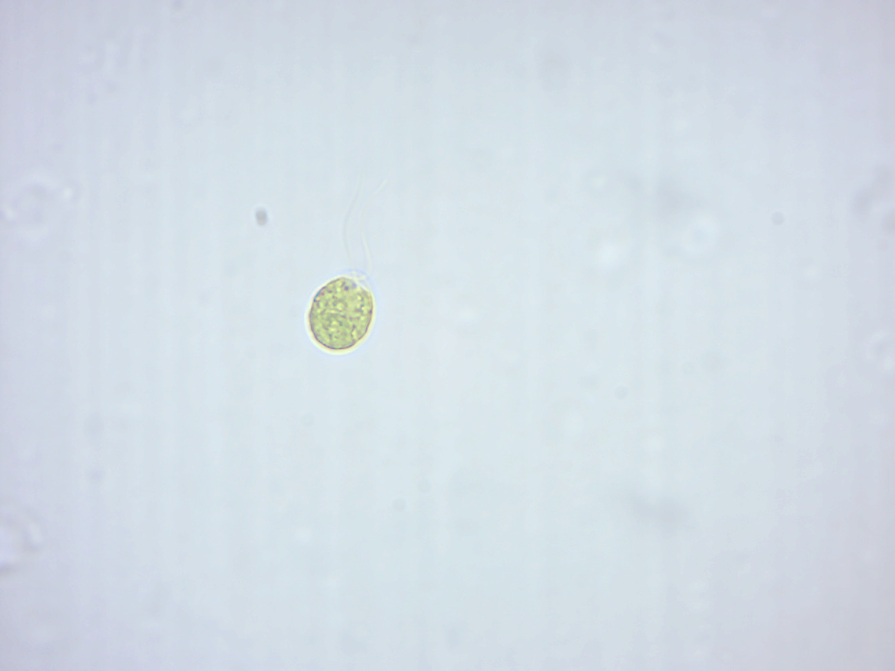
Figure 14.v: Chlamydomonas. Note the flagella.
Gymnodinium
Gymnodinium is a genus of dinoflagellates It is one of the few naked dinoflagellates, or species lacking armor (cellulosic plates). The dinoflagellates (Greek dinos "whirling" and Latin flagellum "whip, scourge") are a large grouping of flagellate eukaryotes that institute the phylum Dinoflagellata. Near are marine plankton, but they are common in freshwater habitats, besides. Their populations are distributed depending on temperature, salinity, or depth. Many dinoflagellates are known to exist photosynthetic, but a large fraction of these are in fact mixotrophic, combining photosynthesis with ingestion of casualty (phagotrophy). In terms of number of species, dinoflagellates class one of the largest groups of marine eukaryotes, although this group is essentially smaller than the diatoms. Some species are endosymbionts of marine animals and play an of import part in the biology of coral reefs. Other dinoflagellates are unpigmented predators on other protozoa, and a few forms are parasitic.
Pandorina
Pandorina (Figure xiv.6) is a genus of green algae composed of 8, 16, or sometimes 32 cells, held together at their bases to grade a sack globular colony surrounded by mucilage. The cells are ovoid or slightly narrowed at one cease to appear keystone- or pear-shaped. Each prison cell has 2 flagella with two contractile vacuoles at their base, an eyespot, and a large loving cup-shaped chloroplast with at to the lowest degree one pyrenoid. The colonies co-ordinate their flagellar movement to create a rolling, swimming motion. Pandorina shows the ancestry of the colony polarity and differentiation seen in Volvox since the inductive cells have larger eyespots. Asexual reproduction is by simultaneous division of all cells of the colony to form autocolonies that are liberated past a gelatinization of the colonial envelope. Sexual reproduction occurs by partitioning of each cell of the colony into xvi-32 zoogametes. Zoogametes bear witness indications of heterogamy, a slight difference in the size and motility of the pairs that fuse to class the polish walled zygote.
Effigy fourteen.vi: Pandorina.
Volvox
Volvox (Effigy 14.seven) is a genus of freshwater algae constitute in ponds and ditches, even in shallow puddles. It forms spherical colonies of up to l,000 cells that were first reported by Antonie van Leeuwenhoek in 1700. Volvox diverged from unicellular ancestors approximately 200 million years agone. Each mature Volvox colony is equanimous of upwards to thousands of cells from two differentiated cell types: numerous flagellate somatic cells and a smaller number of germ cells lacking in soma that are embedded in the surface of a hollow sphere or coenobium containing an extracellular matrix made of glycoproteins. Developed somatic cells contain a single layer with the flagella facing outward. The cells swim in a coordinated fashion, with distinct anterior and posterior poles. The cells have anterior eyespots that enable the colony to swim towards light. An asexual colony includes both somatic (vegetative) cells, which do not reproduce, and large, non-motile gonidia in the interior, which produce new colonies through repeated division. In sexual reproduction two types of gametes are produced. Volvox species can be monoecious or dioecious. Male colonies release numerous sperm packets, while in female person colonies single cells enlarge to become oogametes, or eggs. Volvox is facultatively sexual and can reproduce both sexually and asexually. The switch from asexual to sexual reproduction can exist triggered by environmental conditions and by the product of a sex activity-inducing pheremone. Desiccation-resistant diploid zygotes are produced post-obit successful fertilization.
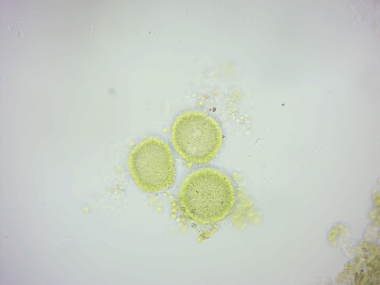
Figure 14.7: Volvox.
Oedogonium
Oedogonium (Figure 14.8) is a genus of filamentous greenish algae, with unbranched filaments that are one jail cell thick. Oedogonium can be free-floating, though information technology is usually attached to aquatic plants by a holdfast. It appears light-green and inhabits calm, fresh h2o. Oedogonium tin can reproduce asexually past fragmentation of the filaments, through some other types of non-motile spores, and also through zoospores, which have many flagella. These develop in a zoosporangium cell, 1 zoospore per zoosporangium. After settling and losing its flagella, a zoospore grows into a filament. Oedogonium tin can too reproduce sexually. Its sexual life cycle is haplontic, i.e., the zygote undergoes meiosis. Antheridia produce and release sperm, and oogonia produce and release an egg,. The egg and sperm then fuse and form a zygote which is diploid (2n). The zygote then undergoes meiosis to produce the filamentous green alga which is haploid (1n).
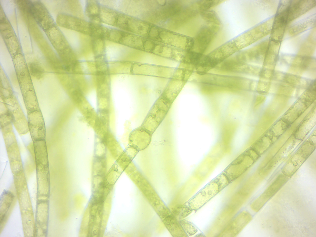
Figure 14.eight: Oedogonium.
Spirogyra
Spirogyra (Figure 14.9; common names include h2o silk, mermaid's tresses, and coating weed) is a genus of filamentous chlorophyte greenish algae of the lodge Zygnematales, named for the helical or spiral organization of the chloroplasts that is diagnostic of the genus. It is commonly constitute in freshwater areas, and in that location are more than 400 species of Spirogyra in the world. Spirogyra measures approximately 10 to 100 μm in width and may abound to several centimeters in length. Spirogyra can reproduce both sexually and asexually. In vegetative reproduction, fragmentation takes place, and Spirogyra simply undergoes the intercalary mitosis to form new filaments. Sexual Reproduction is of two types: one. Scalariform conjugation requires association of two different filaments lined side by side either partially or throughout their length. One jail cell each from contrary lined filaments emits tubular protuberances known as conjugation tubes, which elongate and fuse, to make a passage called the conjugation canal. The cytoplasm of the prison cell interim as the male travels through this tube and fuses with the female person cytoplasm, and the gametes fuse to form a zygospore. ii. In lateral conjugation, gametes are formed in a single filament. Two adjoining cells about the mutual transverse wall give out protuberances known as conjugation tubes, which further class the conjugation canal upon contact. The male cytoplasm migrates through the conjugation canal, fusing with the female. The rest of the process proceeds equally in scalariform conjugation. The essential difference is that scalariform conjugation occurs between ii filaments and lateral conjugation occurs between two next cells on the same filament.
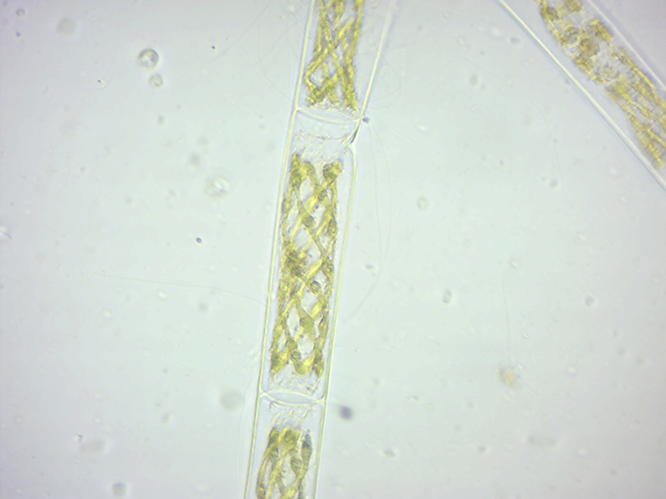
Figure 14.9: Spirogyra.
View Prepared Slides
Amoeba proteus (Figure xiv.10)
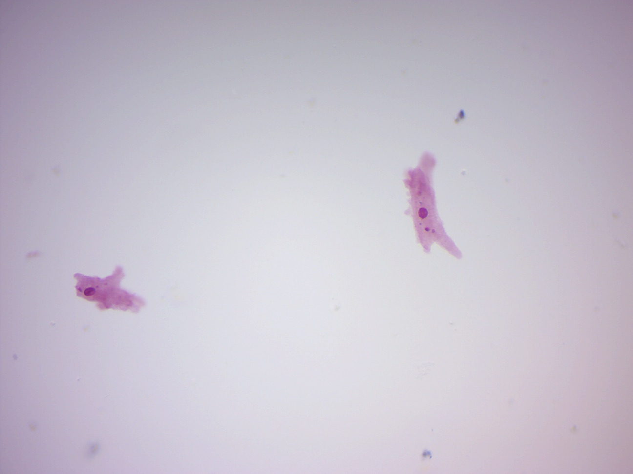
Figure 14.10: Amoeba proteus.
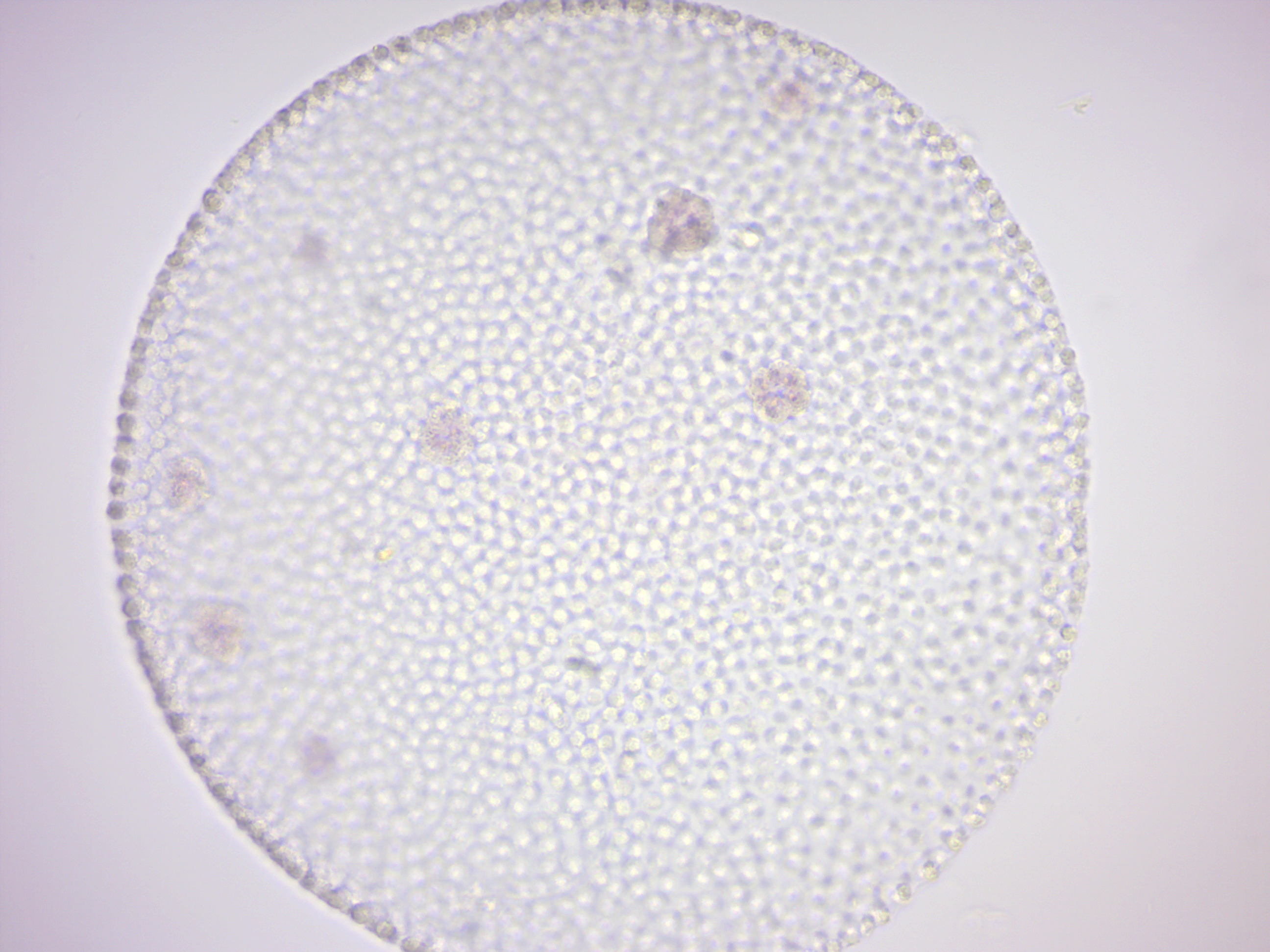
Paramecium 4 types of protista (Figure fourteen.xi)
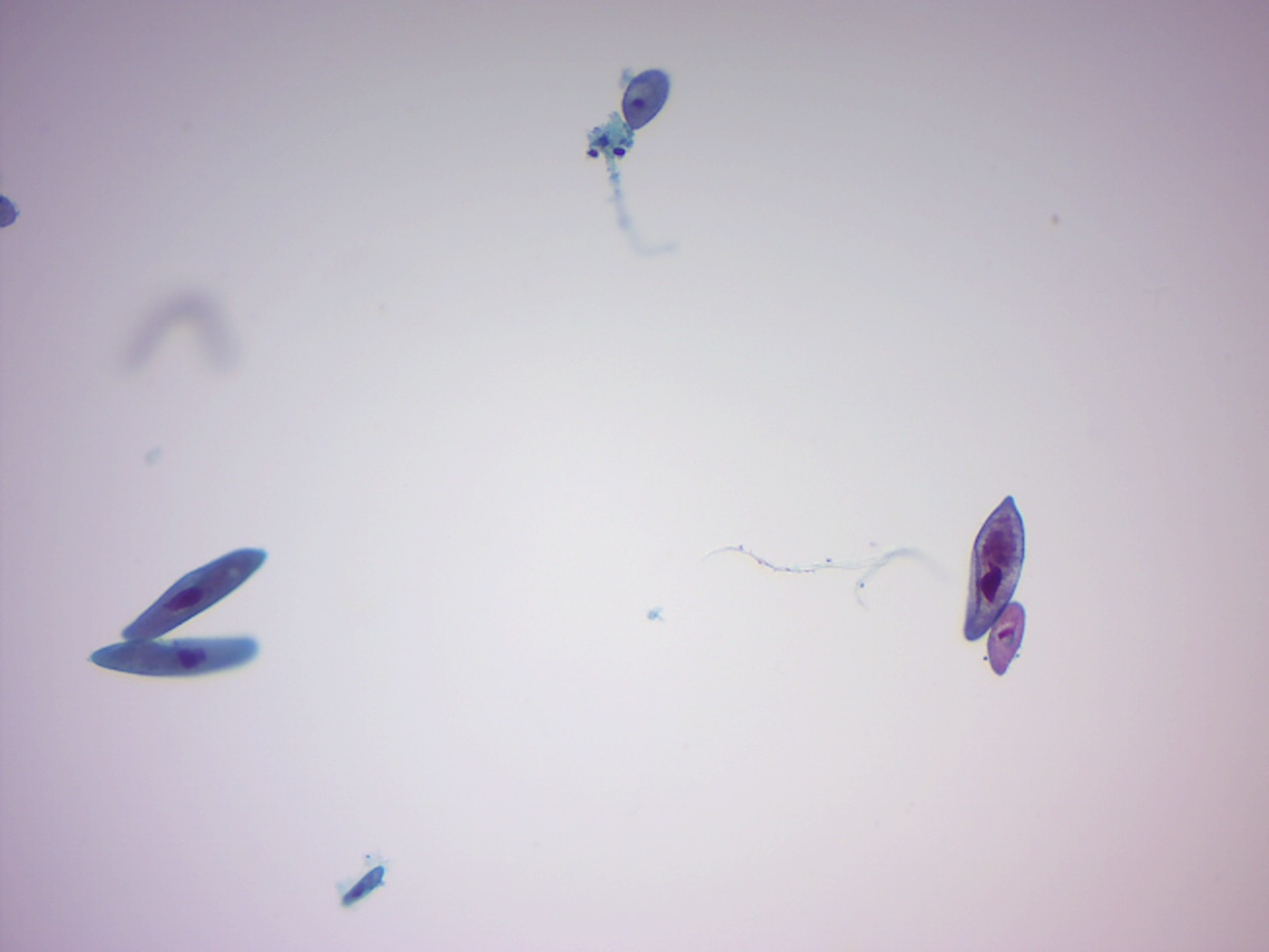
Figure 14.eleven: Paramecia and other protists.
Paramecium caudatum (Effigy 14.12)
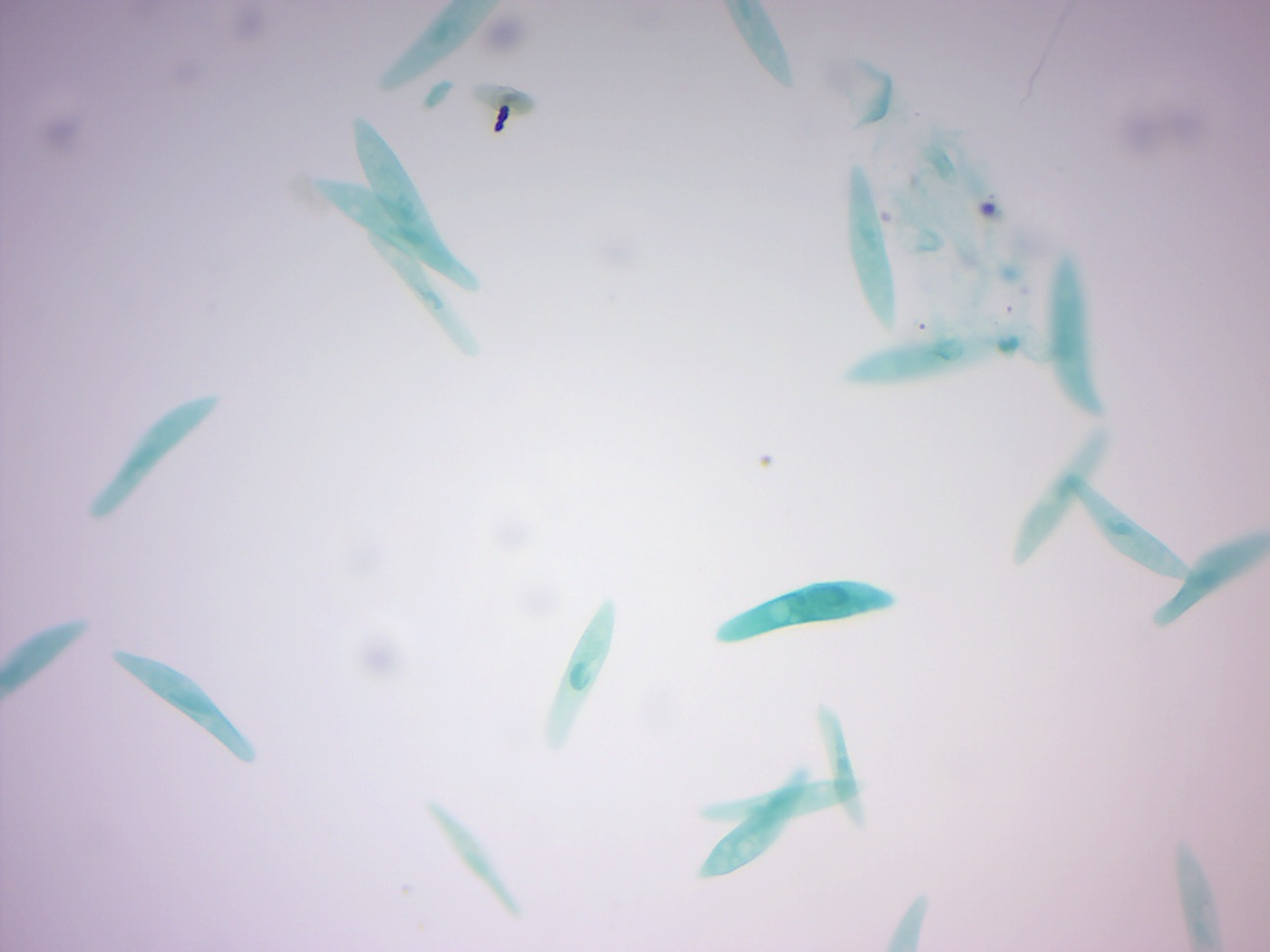
Figure 14.12: Paramecium.
Paramecium in conjugation (Effigy 14.13)
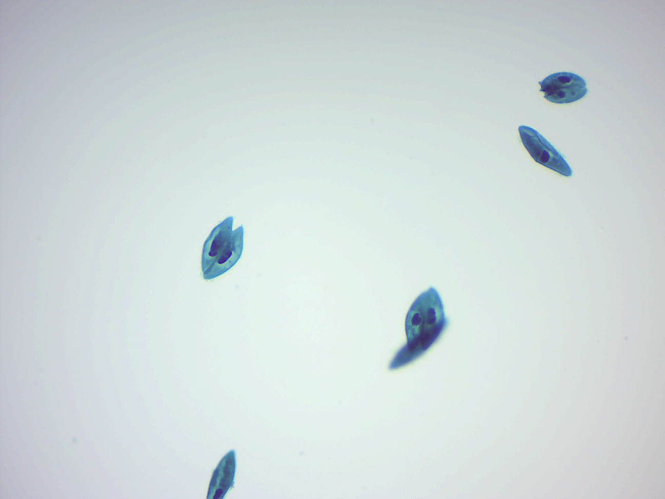
Effigy 14.13: Paramecium in conjugation.
Euglena (Figure xiv.fourteen)
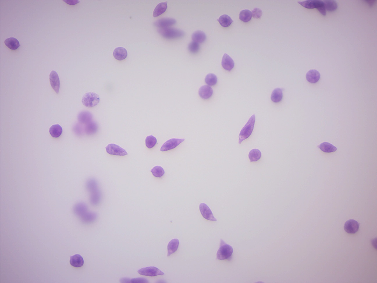
Figure 14.fourteen: Euglena.
Dinoflagellate (Figure xiv.15)
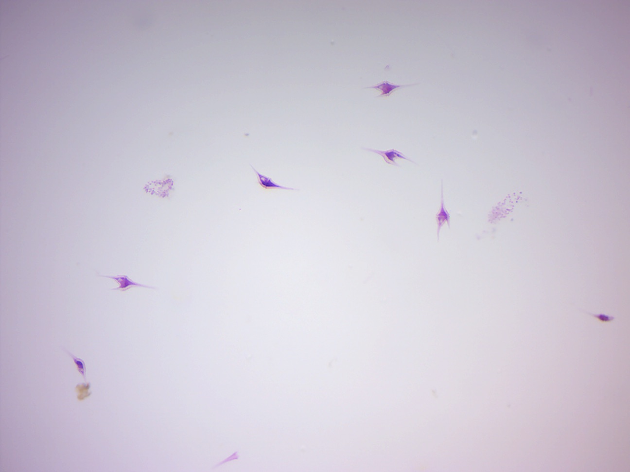
Figure 14.15: Dinoflagellates.
Ceratium (Figure 14.xvi)
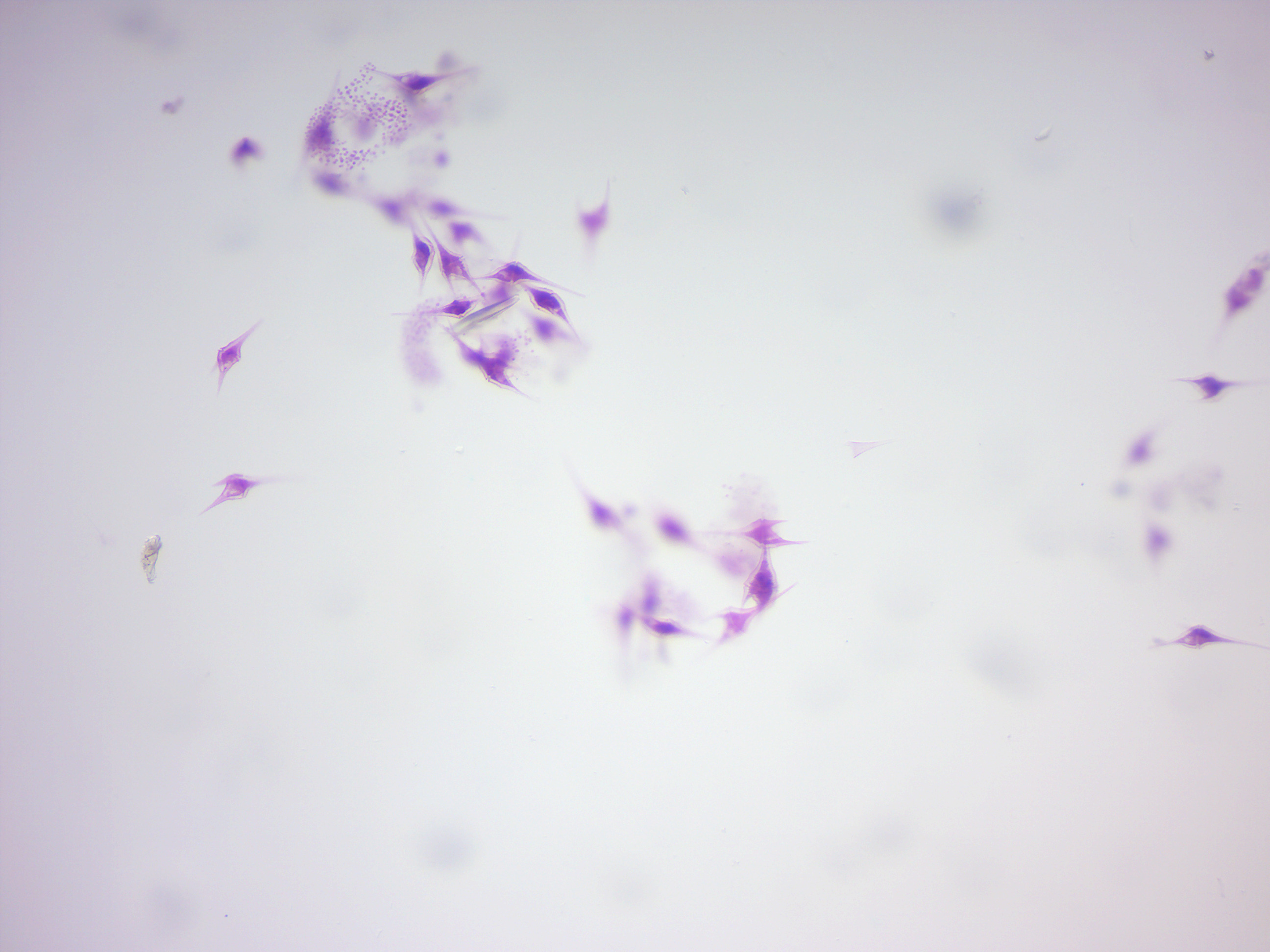
Figure 14.sixteen: Ceratium, a dinoflagellate.
Peridinium (Effigy fourteen.17)
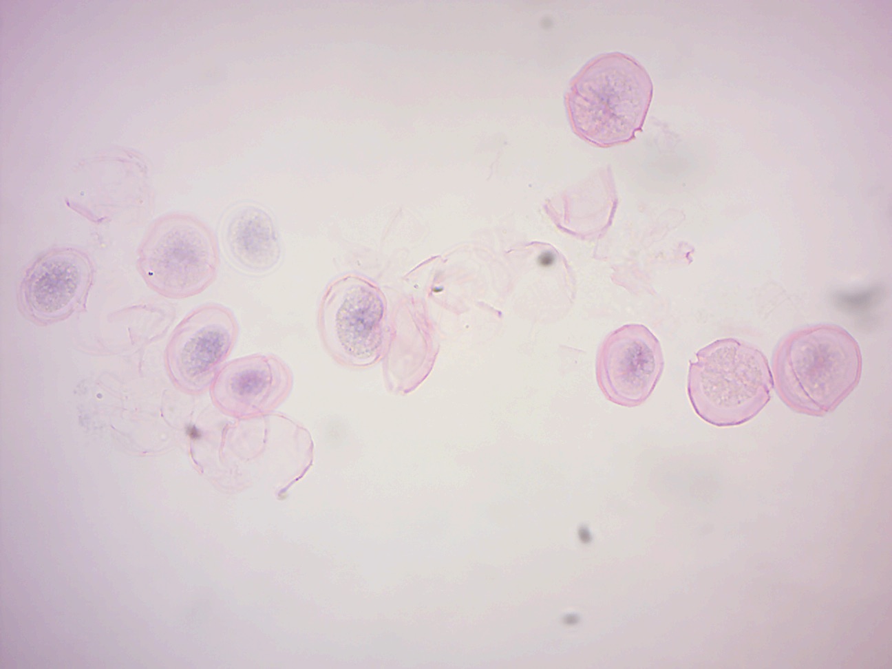
Figure 14.17: Peridinium, a dinoflagellate.
Foraminifera
Foraminifera (Effigy 14.18; Latin meaning hole bearers; informally called "forams") are members of a phylum or course of amoeboid protists characterized by: streaming granular ectoplasm for communicable food and other uses; and commonly an external shell (called a "test") of diverse forms and materials. Most foraminifera are marine, the majority of which live on or within the seafloor sediment (i.east., are benthic), while a smaller variety float in the water column at diverse depths (i.e., are planktonic). These shells are commonly made of calcium carbonate (CaCO3) or agglutinated sediment particles. Over 50,000 species are recognized, both living (10,000) and fossil (40,000).
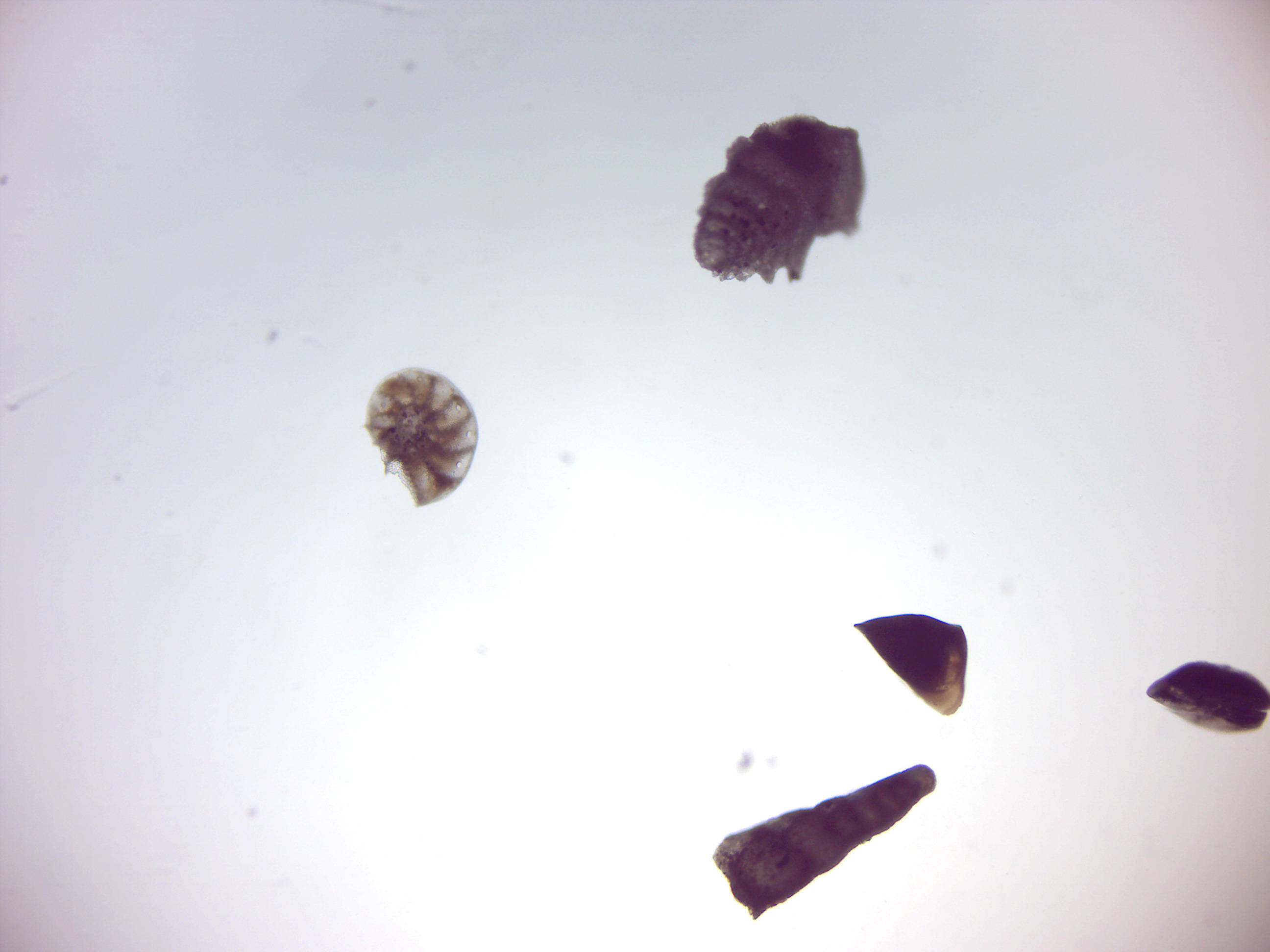
Figure fourteen.18: Foraminifera.
Radiolaria
Radiolaria (Effigy 14.nineteen), also chosen Radiozoa, are protozoa of diameter 0.1-0.2 mm that produce intricate mineral skeletons, typically with a central capsule dividing the prison cell into the inner and outer portions of endoplasm and ectoplasm.The elaborate mineral skeleton is usually made of silica. They are found equally zooplankton throughout the ocean, and their skeletal remains make upwardly a large role of the cover of the ocean flooring equally siliceous ooze.
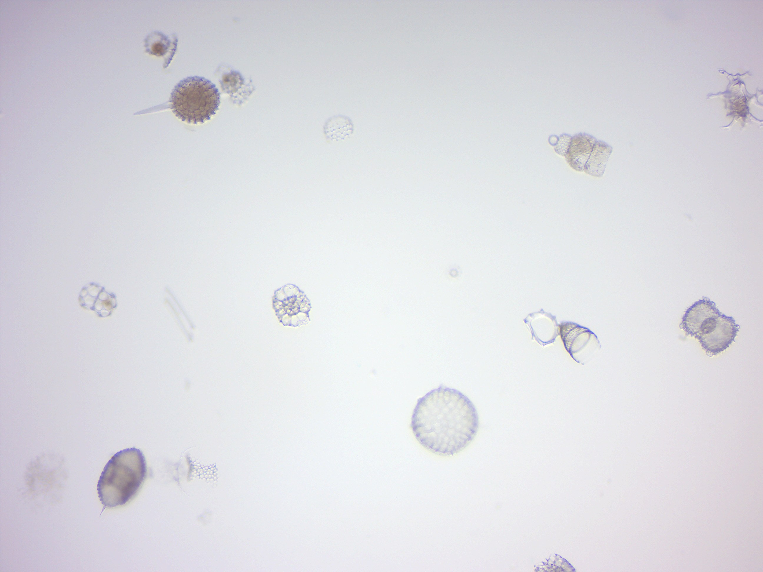
Figure 14.nineteen: Radiolaria.
Diatoms
Diatoms (Figure xiv.xx) are a major group of microalgae and are among the well-nigh mutual types of phytoplankton. Diatoms are producers within the nutrient chain. A unique feature of diatom cells is that they are enclosed within a cell wall made of silica (hydrated silicon dioxide) called a frustule. These frustules show a wide diversity in class, but are normally almost bilaterally symmetrical, hence the group name. These shells are used past humans as diatomaceous earth, also known as diatomite. Fossil show suggests that they originated during, or before, the early on Jurassic period. Merely male person gametes of axial diatoms are capable of move by means of flagella. Diatom communities are a popular tool for monitoring environmental conditions, past and present, and are ordinarily used in studies of water quality.
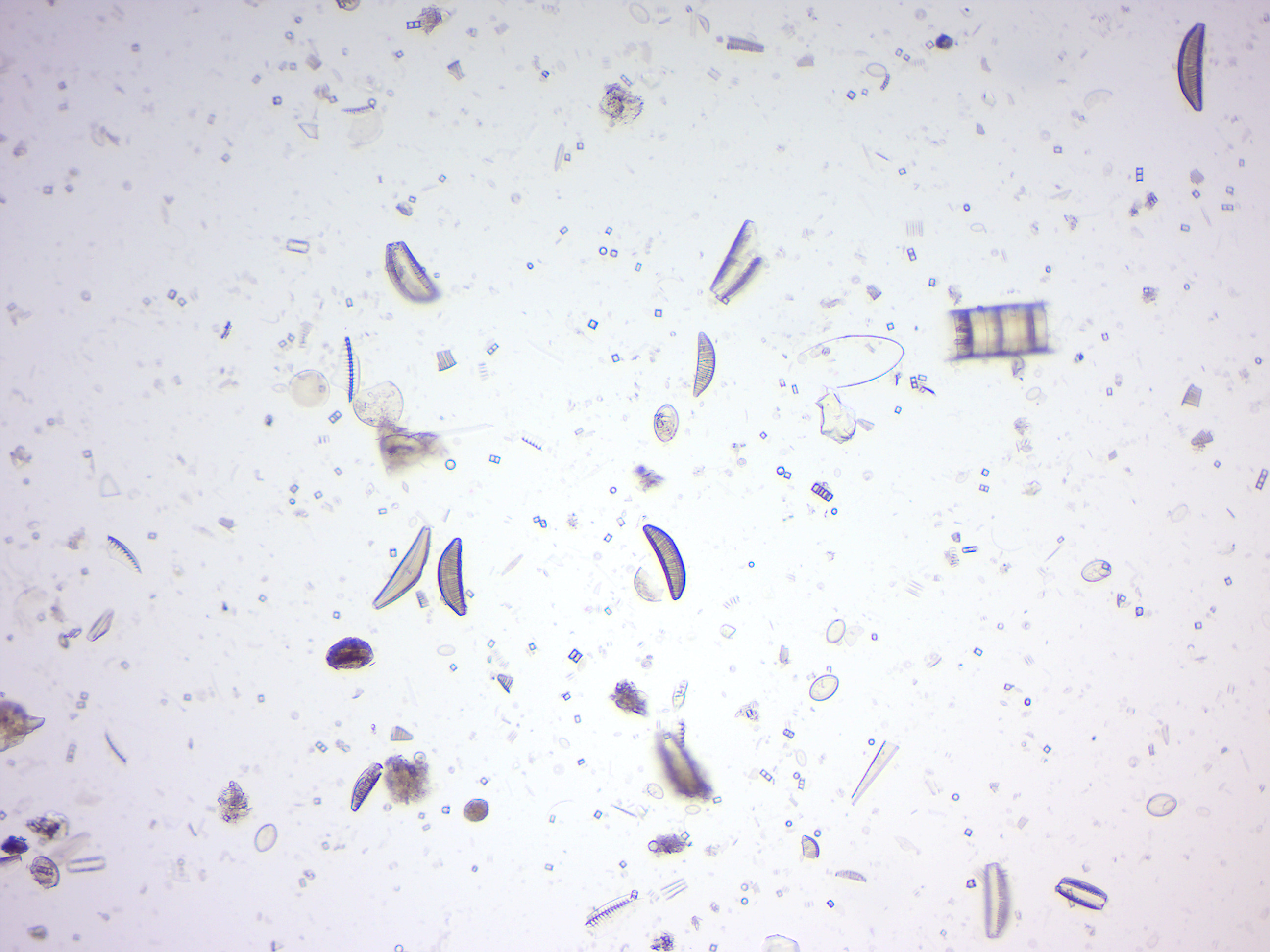
Figure 14.20: Diatomes.
Trypanosoma cruzi and Trypanosoma brucei gambiense
Trypanosoma cruzi is a species of parasitic euglenoids. Amongst the protozoa, the trypanosomes characteristically bore tissue in another organism and feed on claret (primarily) and also lymph. This behaviour causes illness or the likelihood of disease that varies with the organism: for example, trypanosomiasis in humans (Chagas affliction in South America). Parasites need a host body and the haematophagous insect triatomine (descriptions "assassin bug", "cone-nose issues", and "kissing problems") is the major vector in accord with a mechanism of infection. The triatomine likes the nests of vertebrate animals for shelter, where it bites and sucks blood for food. Individual triatomines infected with protozoa from other contact with animals transmit trypanosomes when the triatomine deposits its faeces on the host'south skin surface and and so bites. Penetration of the infected faeces is further facilitated by the scratching of the bite area by the human or animal host.
Trypanosoma brucei (Effigy xiv.21) is a species of parasitic kinetoplastid belonging to the genus Trypanosoma. The parasite is the crusade of a vector-borne affliction of vertebrate animals, including humans, carried by genera of tsetse fly in sub-Saharan Africa. In humans T. brucei causes African trypanosomiasis, or sleeping sickness. In animals it causes animal trypanosomiasis, also called nagana in cattle and horses. T. brucei has traditionally been grouped into iii subspecies: T. b. brucei, T. b. gambiense and T. b. rhodesiense. The first is a parasite of non-human vertebrates, while the latter two are the known parasites of humans.
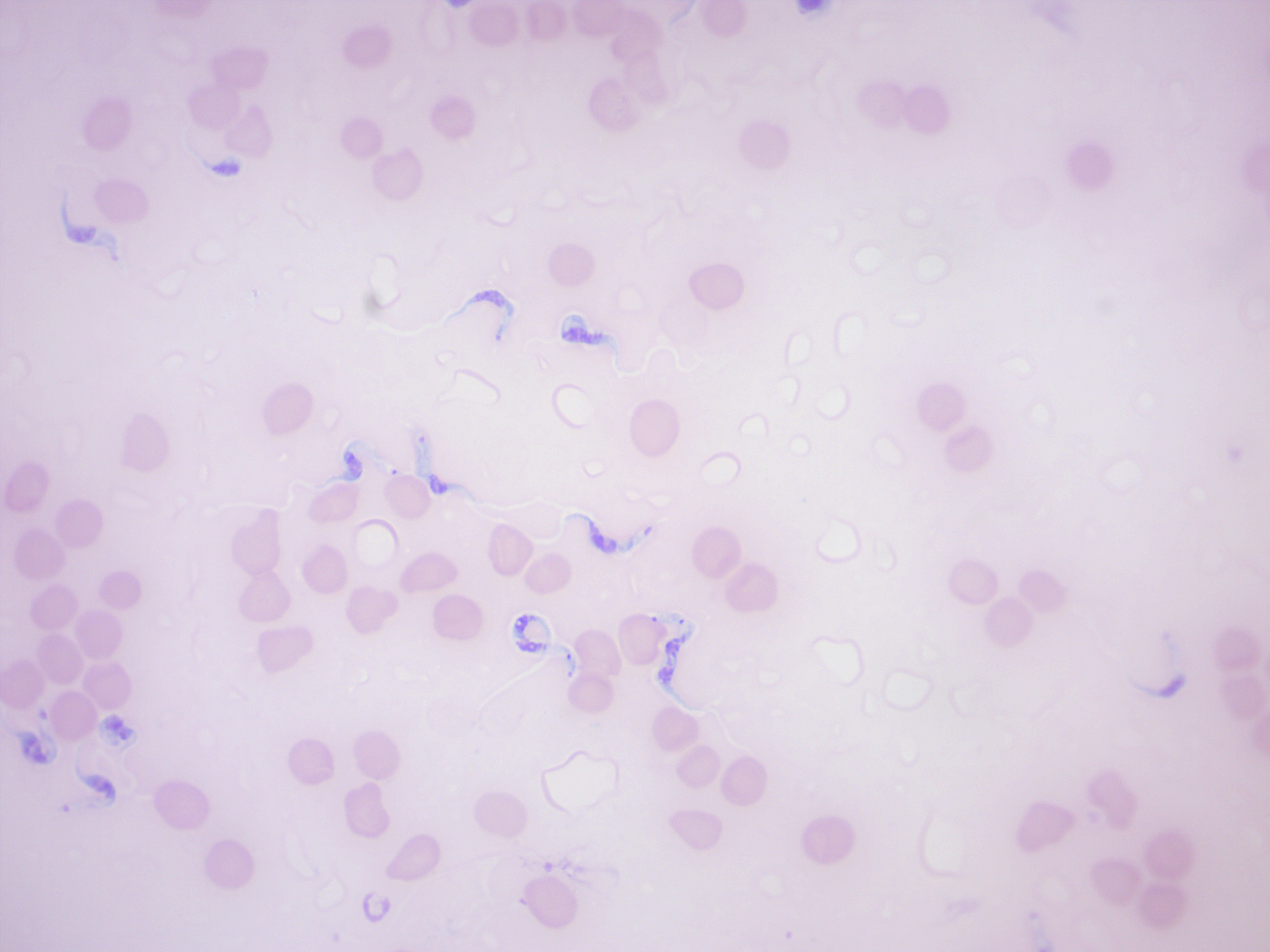
Figure 14.21: Trypanosoma brucei gambiense among carmine claret cells.
Plasmodium vivax
Plasmodium vivax (Figure xiv.22) is a protozoal parasite and a human pathogen. This parasite is the almost frequent and widely distributed cause of recurring (benign tertian) malaria, P. vivax is 1 of the 5 species of malaria parasites that commonly infect humans. Although information technology is less virulent than Plasmodium falciparum, the deadliest of the five human being malaria parasites, P. vivax malaria infections can lead to severe disease and decease, oft due to a pathologically enlarged spleen. P. vivax is carried by the female Anopheles mosquito, since it is just the female of the species that bites.
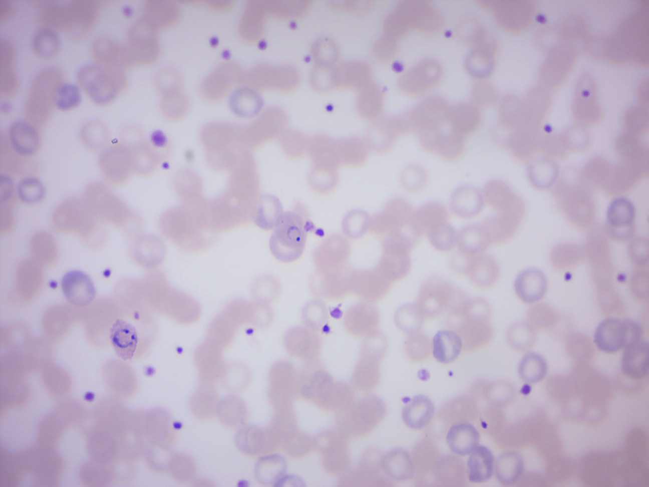
Figure xiv.22: Plasmodium vivax merozoites and trophozoites (ring stage).
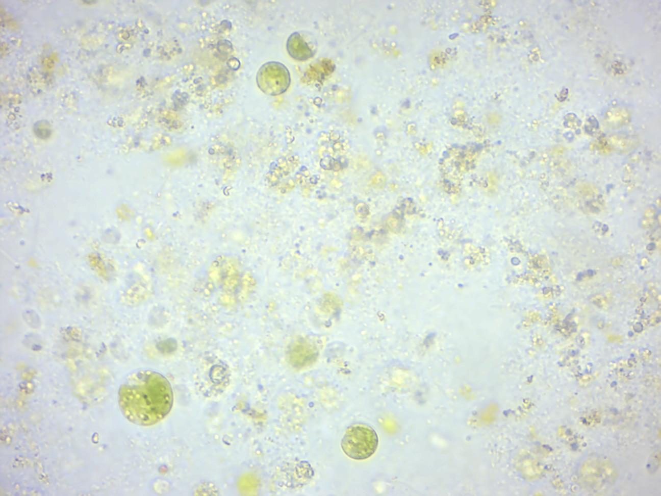
Mixed green algae (Figure 14.23)
Effigy 14.23: Various green algae.
Chlamydomonas (Effigy xiv.24)
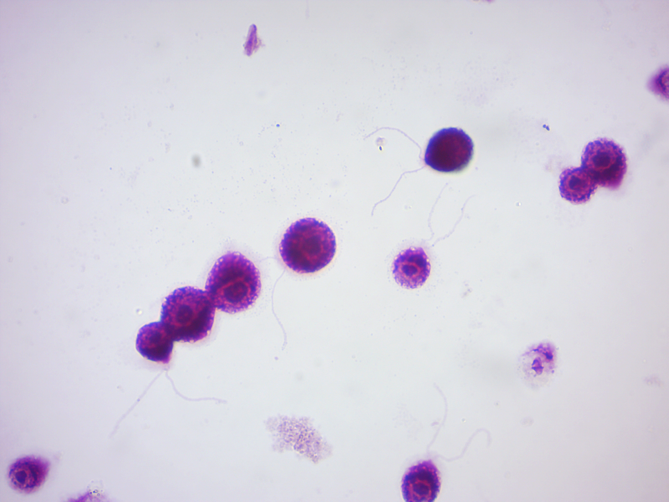
Figure 14.24: Chlamydomonas. Notation the flagella.
Pandorina (Figure 14.25)
Figure fourteen.25: Pandorina.
Volvox (Figure 14.26)
Effigy xiv.26: Volvox.
Volvox sexual stages (Effigy 14.27)
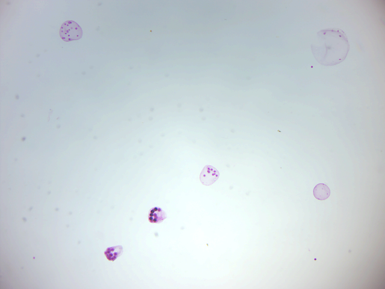
Figure fourteen.27: Volvox sexual stages.
Spirogyra (Effigy 14.28)
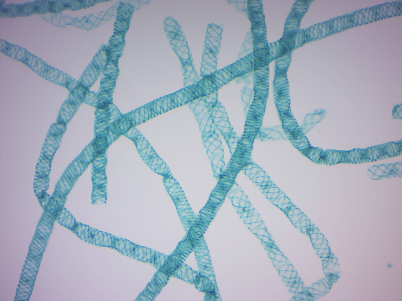
Figure 14.28: Spirogyra.
Oedogonium zoospores (Figure xiv.29
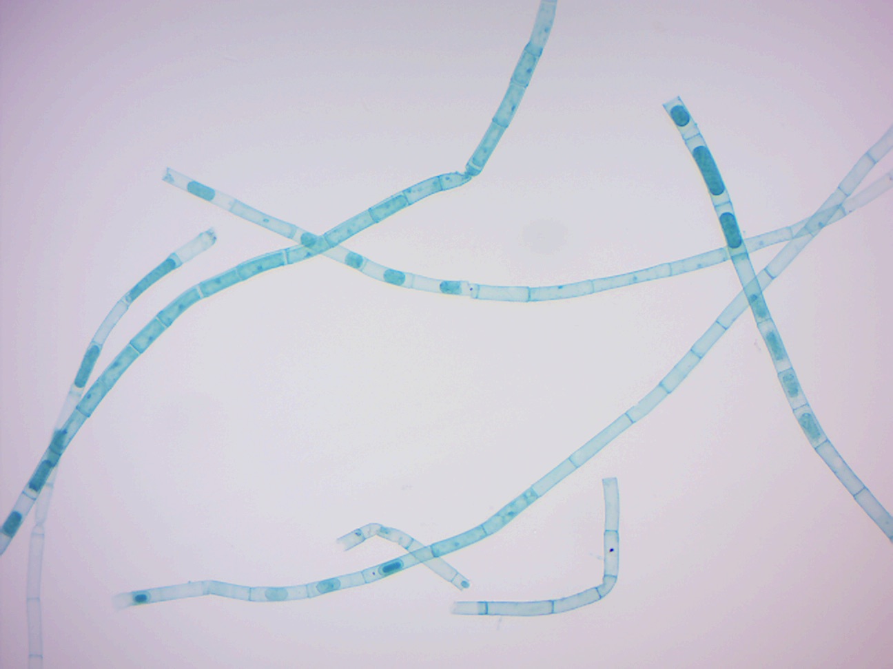
Figure xiv.29: Oedogonium zoospores.
Oedogonium macrandous (Figure 14.xxx)
Effigy 14.30: Oedogonium.
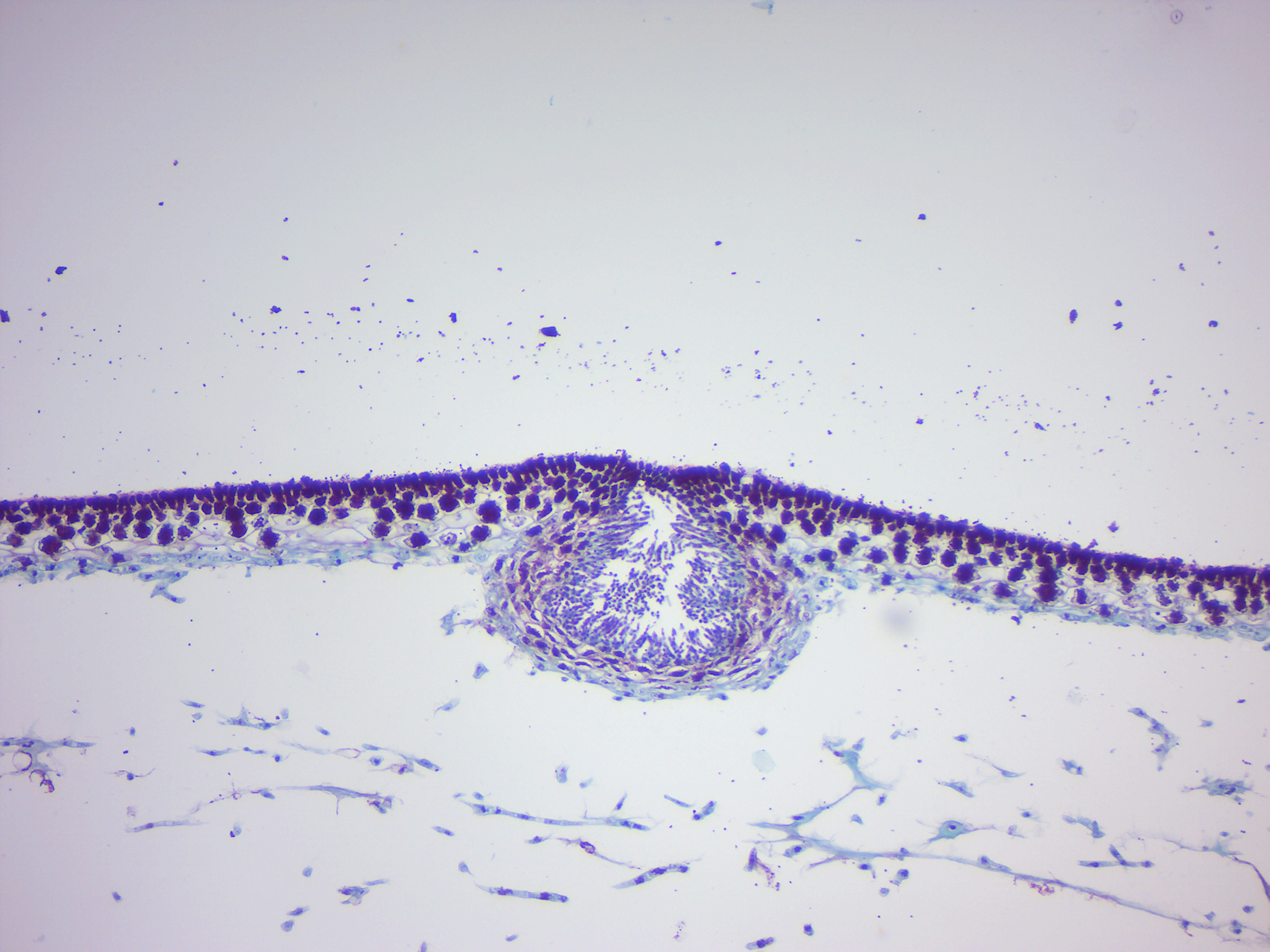
Fucus male and female conceptacle
Fucus is a genus of chocolate-brown algae found in the intertidal zones of rocky seashores almost throughout the world. It has a relatively simple life wheel and produce only one blazon of thallus which grows to a maximum size of ii m. The thallus is perennial with an irregular or disc-shaped holdfast or with haptera. The erect portion of the thallus is dichotomous or subpinnately branched, flattened and with a distinct midrib. Gas-filled pneumatocysts (air-vesicles) are present in pairs in some species, i on either side of the midrib. The gametangia develop in conceptacles embedded in receptacles in the apices of the final branches. They may exist monoecious or dioecious. Fertile cavities, the conceptacles, containing the reproductive cells are immersed in the receptacles nearly the ends of the branches. After meiosis oogonia and antheridia are produced and released, fertilisation follows and the zygote develops directly into the diploid institute. It may be considered to exist analogous to the life cycle of the flowering plant, but in algae the oogonia are released and fertilised in the sea while in flowering plants the ovules are fertilised while attached to the parent plant and then released every bit a seed.
Fucus male conceptacle (Figure 14.31)
Effigy 14.31: Fucus male person conceptacle
Fucus female conceptacle (Effigy 14.32)
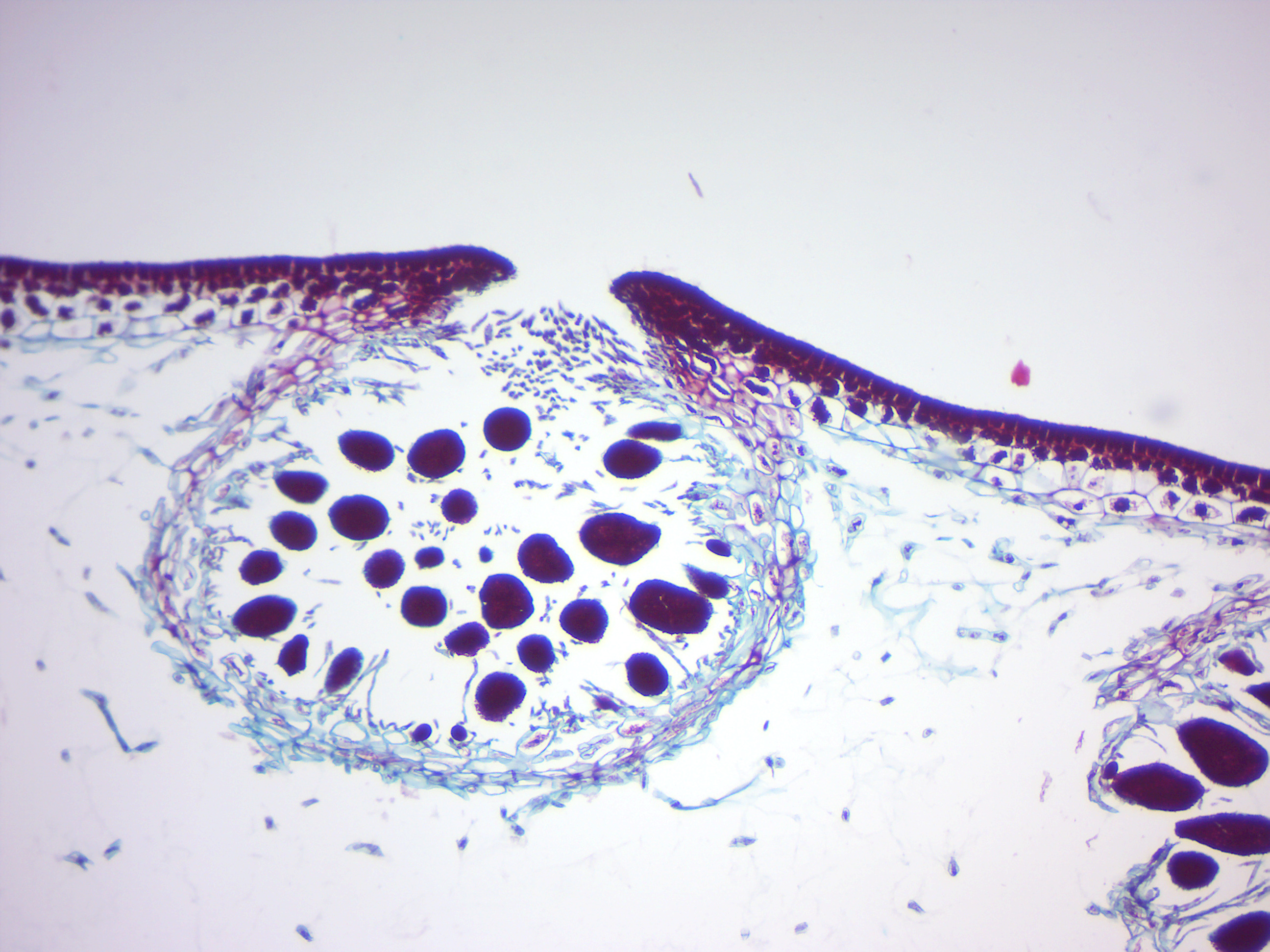
Figure 14.32: Fucus female conceptacle
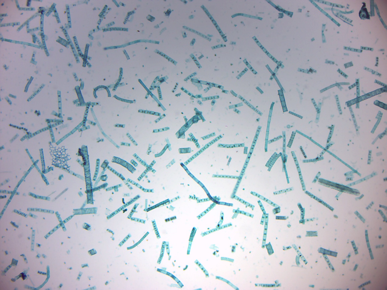
Polysiphonia
Polysiphonia (Effigy 14.33) is a genus of filamentous red algae with most nineteen species on the coasts of the British Isles and nigh 200 species worldwide.
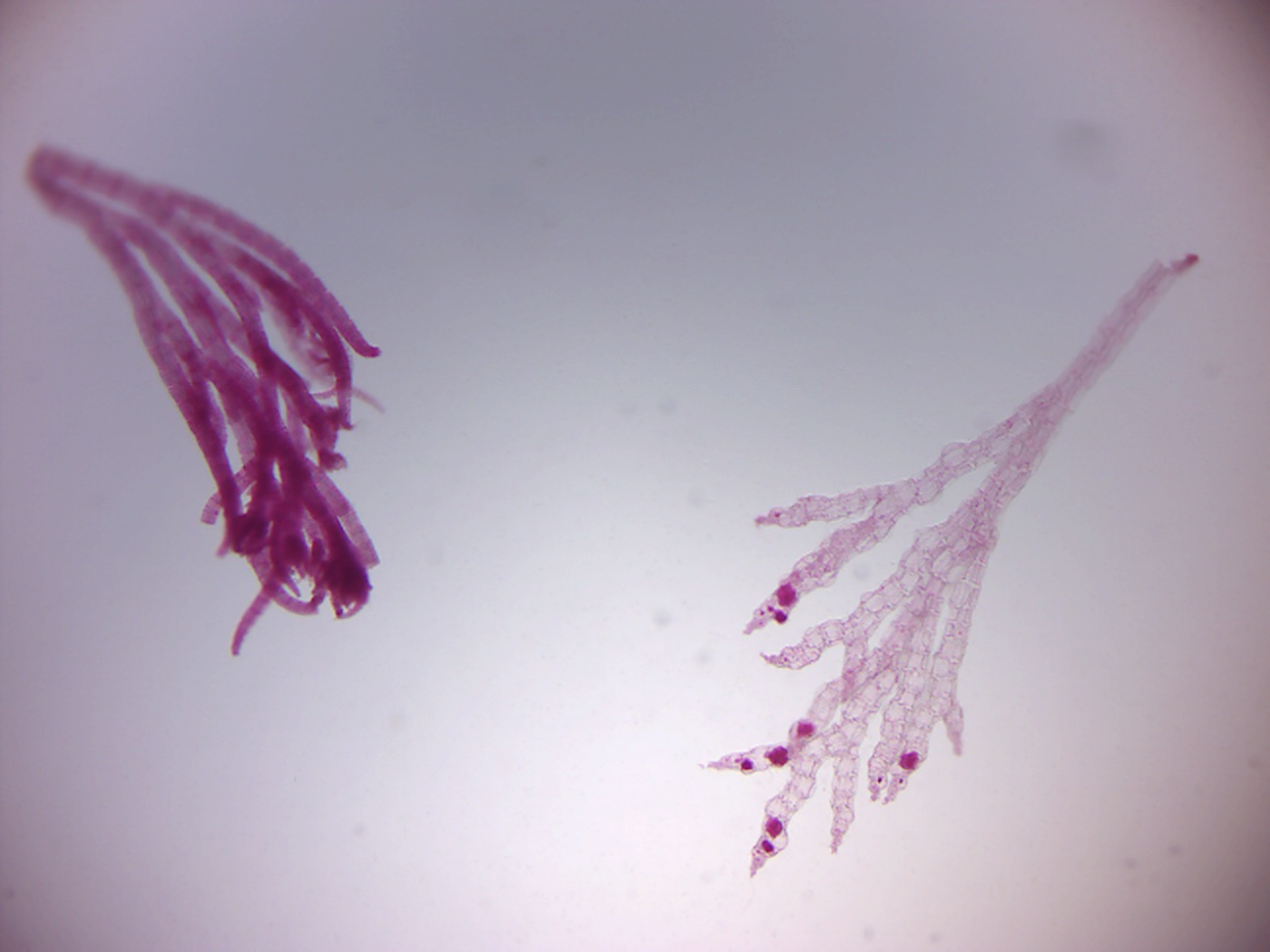
Figure 14.33: Polysiphonia.
Stemonitis
Stemonitis (Effigy xiv.34) is a distinctive genus of slime moulds institute throughout the world (except Antarctica). They are characterized past the tall brown sporangia, supported on slender stalks, which grow in clusters on rotting wood.
Figure 14.34: Stemonitis.
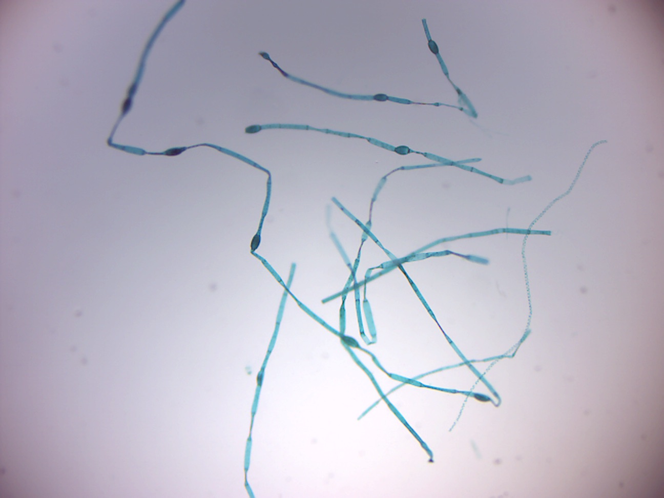
Saprolegnia
Saprolegnia (Figure xiv.35) is both a saprotroph and necrotroph. Typically feeding on waste from fish or other expressionless cells, they will too take advantage of creatures that take been injured. An infection is known equally oomycosis Saprolegnia is tolerant to a wide range of temperature, 3 °C to 33 °C, merely is more prevalent in lower temperatures. While it is found nearly ofttimes in freshwater, it will also tolerate brackish water and even moist soil. Saprolegnia filaments (hyphae) are long with rounded ends, containing the zoospores. Saprolegnia by and large travels in colonies consisting of one or more species. They first course a mass of individual hyphae. When the mass of hyphae grows large enough in size to be seen without use of a microscope, it can be called a mycelium.

Figure 14.35: Saprolegnia.
It has a diploid life bike which includes both sexual and asexual reproduction. In the asexual phase, a spore of Saprolegnia releases zoospores. Within a few minutes, this zoospore will encyst, germinate and release another zoospore. This second zoospore has a longer cycle during which almost dispersal happens; it will proceed to encyst and release a new spore in a process called polyplanetism until information technology finds a suitable substrate. When a suitable medium is located, the hairs surrounding the spore volition lock onto the substrate so that the sexual reproduction phase can start. Information technology is also during this stage of polyplanetism that the Saprolegnia are capable of causing infection; the most pathogenic species have tiny hooks at the stop of their hairs to raise their infectious ability. Once firmly attached, sexual reproduction begins with the production of male person and female person gametangium, antheridia and oogonium respectively. These unite and fuse together via fertilization tubes. The zygote produced is named an oospore.
Review Questions
- What are protists?
- What are ciliata?
- What are flagellata?
- How do amoeba move?
- What are slime molds?
Source: https://myrcc.rcc.mass.edu/ICS/103_lab_manual/protista.html
0 Response to "Lab 6 Review Is Onion Skin Multicellular or Unicellular or Colonial"
Post a Comment